Figure 1:
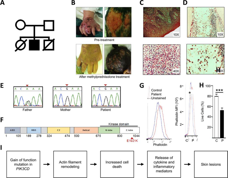
Characterization of patient phenotype. (A) Family pedigree. (B) Inflammatory skin lesions pre- and post-treatment with methylprednisolone. (C) Skin biopsy (hematoxylin & eosin; original magnification 10X and 40X) showing epidermis with hyperparakeratosis, acanthosis, papillomatosis, spongiosis and inflammatory infiltrate composed of neutrophils, lymphocytes and eosinophils. Dermis has mixed inflammatory infiltrate, with numerous eosinophils at magnification 40x. (D) Bone marrow biopsy (hematoxylin & eosin; original magnification 10X and 40X) showing myeloid predominance due to increased mature eosinophils. Bands, metamyelocytes and myelocytes are also observed. (E) Sanger sequencing of PIK3CD c.3061G>A, pGlu1021Lys variant. (F) Linear diagram of PIK3CD. Patient mutation in red. (G) F-actin content assessed by median fluorescent intensity of permeabilized BLCLs stained with phalloidin-FITC. N=2 pts and 2 controls in 2 independent experiments. (H) Cell death in BLCLs from 3 controls and 2 patients, pooled from 3 independent experiments. (I) Proposed mechanism by which dysregulated actin dynamics may contribute to APDS pathology. Columns and bars represent means +/− SEM. *p<0.05, *** p<0.001; Student’s t test.

