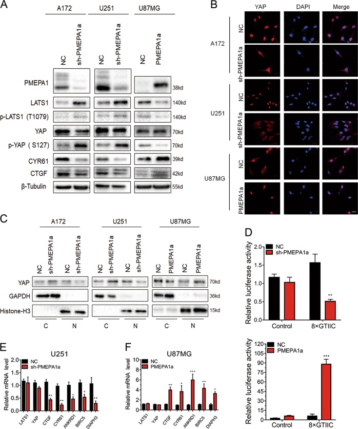
Fig. 4.
PMEPA1a suppresses the Hippo kinase signaling through LATS1. a Western blot analysis to evaluate components in the Hippo kinase pathway downstream of PMEPA1a in lysates prepared from glioma cell lines, which were modified with PMEPA1a or sh-PMEPA1a as indicated. β-Tubulin was used as loading control. b Representative images of immunofluorescence staining for YAP (red) in modified glioma cell lines showing cellular localization. Nuclei are stained with DAPI (blue). Scale bars, 20 µm. c Western blot analysis of cytoplasmic (C) and nuclear (N) fractions prepared from indicated cells. d Luciferase assay for 8xGTIIC-Lux or control reporter constructs indicating YAP-dependent transcriptional activity in modified U87MG and U251 cells. Data are normalized to a Renilla reporter and to the NC group. e, f qRT-PCR analysis of modified U87MG and U251 cells. GAPDH was used as loading control. Data are normalized to the NC group. Student’s t-test: *P < 0.05, **P < 0.01, ***P < 0.001

