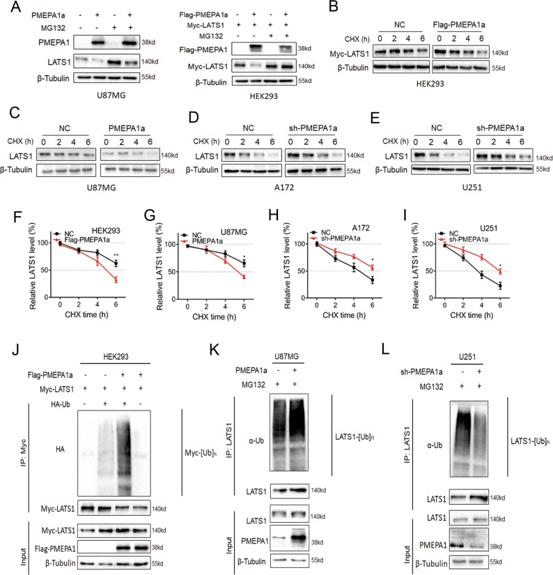
Fig. 5.
PMEPA1a destabilizes LATS1 proteins. a Western blot analysis to evaluate LATS1 levels in U87MG- and HEK293-NC and -PMEPA1a cells after treatment with proteasome inhibitor MG132 (20 µM) for 8 h. β-tubulin was used as the loading control. b–e Western blot analysis of LATS1 protein in modified HEK29, U87MG, A172, and U251 cells treated with cycloheximide (CHX; 25 µg/mL) for the indicated time. (f–i) Line graphs representing LATS1 levels normalized to β-tubulin and to 0 h at the indicated time points from CHX experiments (n = 4). Data are represented as the mean ± SEM. (j–l) Western blot analysis of ubiquitination assays. Cells were transfected with PMEPA1a in HEK293 and U87MG or shRNA (sh-PMPA1a) in U251 cells. Cells were treated with MG132 (20 µM) for 8 h. Student’s t-test: *P < 0.05, **P < 0.01

