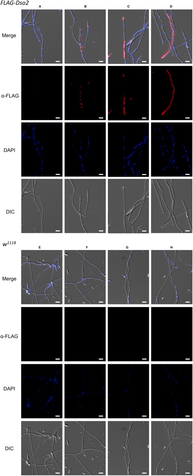
Figure 7.
Immunofluorescence staining of F. oxysporum hyphae. Images showing various staining patterns of hyphae incubated with FLAG-Dso2 (A–D) or w1118 hemolymph (E–H) and then stained with mouse α-FLAG M2 (1:200) and donkey α-mouse Alexa 555 (1:400). DAPI marks fungal DNA. Scale bar is 10 μm. Images were generated as focused images from Z-stacks.

