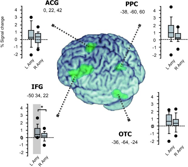
Figure 5.
Functional connectivity with the amygdala in the left-hemisphere network involved in fast emotion word processing. Box plots for each region show the magnitude of group-level inter-regional connectivity with the left and right amygdala ROIs. The four cortical regions identified in the whole-brain SPM (see Figure 3) are surface-rendered on the normalized brain using MRIcron (https://www.nitrc.org/projects/mricron). Significant functional coupling was observed only between the left IFG and the left amygdala (shaded in gray, see section “Results”). Moreover, while all the four regions showed the same trend of greater connectivity with the left amygdala than with the right amygdala, this left-vs.-right difference in amygdala connectivity was significant only for the left IFG (*p = 0.04).

