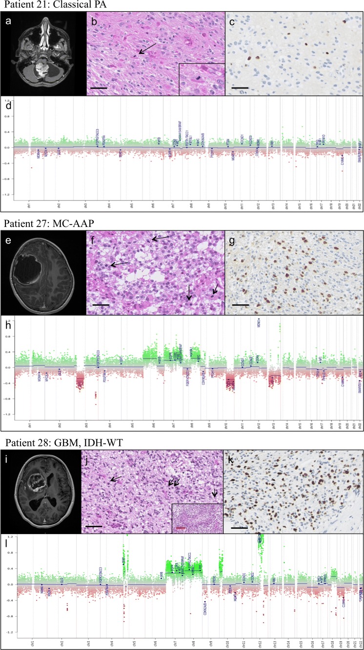
Fig. 3.
Comparative radiological, histological, molecular and CNP characteristics of PA (patient 21), MC-AAP (patient 27), and GBM (patient 28). Patient 21 Classical PA: a MRI: 3D T1 with gadolinium injection, showing a classical cerebellar PA features with both cystic and nodular portions and strong contrast enhancement. b Histological features: biphasic architecture, Rosenthal fibers, eosinophilic granular bodies, and mitosis (see insert and arrowhead), c MIB1 labeling index estimated at 10%, d flat CNP characteristic of classical PA. Patient 27 MC-AAP: e MRI: 3D T1 with gadolinium injection showing a right parietal mostly cystic lesion with a thick, irregular Contrast enhancing border. f Histological features: oligodendroglial-like morphology, numerous mitoses (arrowheads). No Rosenthal fiber or eosinophilic granular bodies were observed, g MIB1 labeling index estimated at 30%, h CNP with many chromosomal gains and losses, as well as focal amplifications, including MDM2. Patient 28 GBM, IDH-WT:i MRI: 3D T1 with gadolinium injection showing a right mass growing from the optic chiasm into the basal ganglia. j Histological features: biphasic morphology with numerous mitoses (arrowheads) and palisading necrosis (see insert), k MIB1 labeling index estimated at 40%, l CNP showing multiple chromosomal gains and losses, including a loss of CDKN2A/B, and focal amplifications of MDM2 and CDK4. Black scale bar corresponds to 50 μm, red scale bar corresponds to 200 μm

