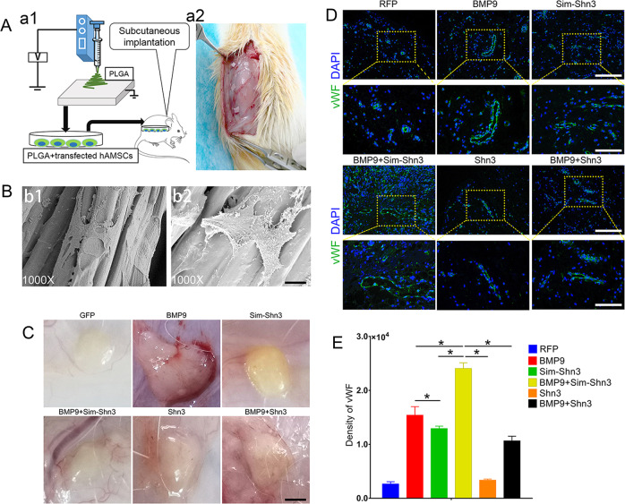
Fig. 7. Effects of Shn3 on subcutaneous vascular invasion of PLGA–hAMSCs composite in vivo.
a Illustrative diagram showing that hAMSCs treated as experimental design seeded on the electro-spun PLGA scaffolds (a1) were implanted into the dorsal subcutaneous space of the mice and harvested for analysis after 5 weeks (a2). b The scanning electron microscopy was adopted to detect the PLGA (b1) and cells cultured on PLGA scaffold (b2) (magnification ×1000, scale bar = 200 μm). c The macromorphological observation of PLGA implants located on subcutaneous tissue at 5 weeks. d, e Immunofluorescence staining (d) and its quantification analysis (e) were performed to detect the vWF expression (green) in PLGA scaffold subcutaneously (magnification of up images = ×200, of down images ×400, scale bar = 50 μm). *P < 0.05.

