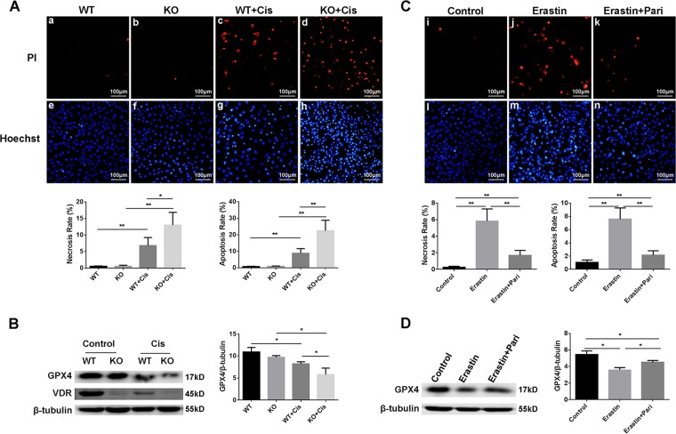
Fig. 8. Worsened cell injury in VDR deficient HK-2 cell induced by cisplatin (A and B), Paricalcitol alleviated Erastin induced cell death in HK-2 cells (C and D).
A PI (a–d) and Hoechst (e–h) staining of wild type and VDR-KO cells after cisplatin treated, and statistical analysis of necrosis and apoptosis rates respectively of each group of cells. Scale bar = 100 μm. B Western blots analysis of GPX4 and VDR in cells from these four groups. The ratio of the optical density of GPX4 to β-tubulin was statistically analyzed. C PI (i–k) and Hoechst (l–n) staining of cells from control, erastin, erastin + paricalcitol groups, respectively followed with statistical analysis of necrosis and apoptosis rates of each group of cells. Scale bar = 100 μm. D Western blots analysis of GPX4 in cells from these three groups. The ratio of the optical density of GPX4 to β-tubulin was statistically analyzed. The data are presented as the mean ± SD, *P < 0.05, **P < 0.01. n = 3.

