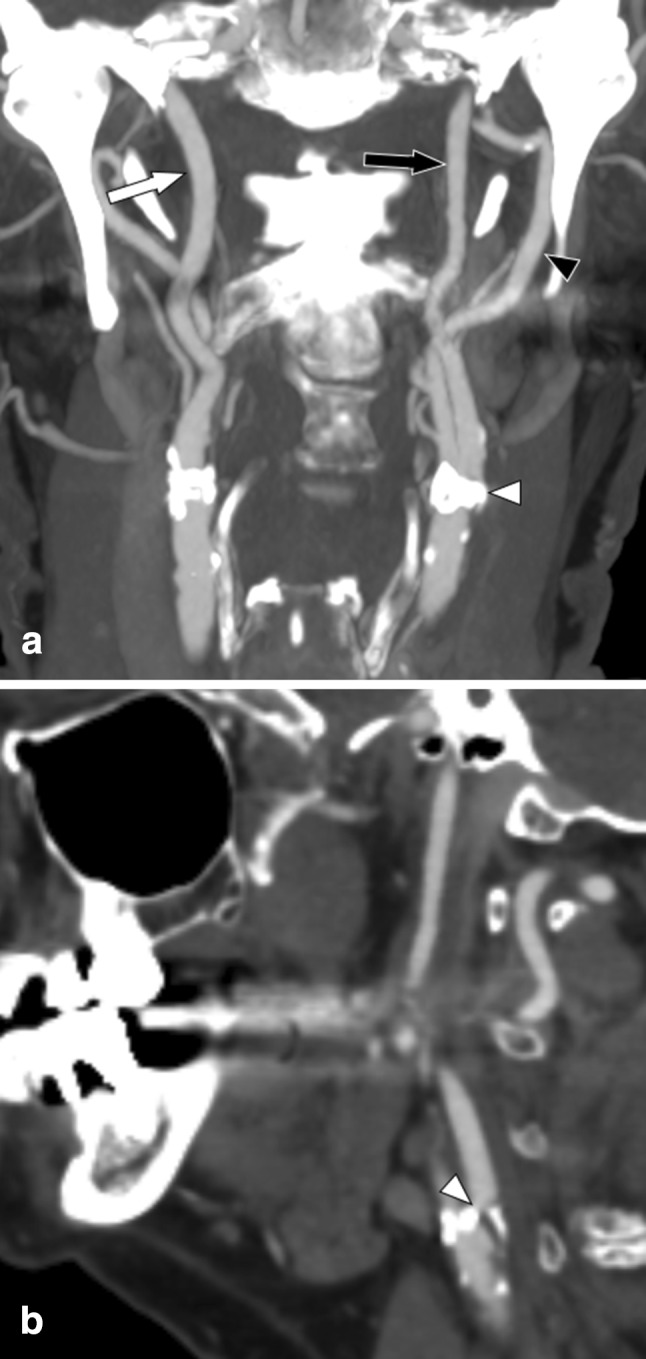
Fig. 1.

Left-sided symptomatic near-occlusion without full collapse; patient suffered a recurrent ipsilateral stroke 5 days after the exam. a Coronal view. Left distal ICA (black arrow, 3.3 mm) is smaller than right distal ICA (white arrow, 4.4 mm) and similar to left ECA (black arrowhead, 3.3 mm). Stenosis is hard to visualize (white arrowhead). b Sagittal view. Stenosis (white arrowhead) somewhat better visualized; lumen at stenosis is tight though but still difficult to assess. Axial source images (not displayed here) are usually most reliable to assess stenosis severity and with other features. A severe stenosis causing flow reduction was the most reasonable explanation of the small distal left ICA, interpreted as near-occlusion
