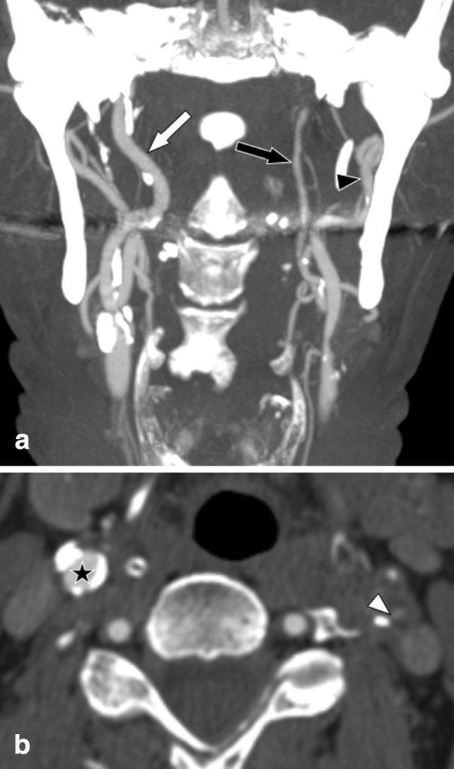
Fig. 2.

Left-sided symptomatic near-occlusion with full collapse; patient suffered a recurrent ipsilateral stroke 7 days after the exam. Left-sided symptomatic near-occlusion with full collapse. a Coronal view. Left distal ICA (black arrow, 1.2 mm) is clearly smaller than right distal ICA (white arrow, 3.7 mm) and smaller than left ECA (black arrowhead, 3.8 mm). Stenosis not seen in this projection, as the lumen was out-of-plane and so small on the image. b Axial view of the stenosis (white arrowhead, 0.8 mm). No relevant stenosis in left ICA (black start)
