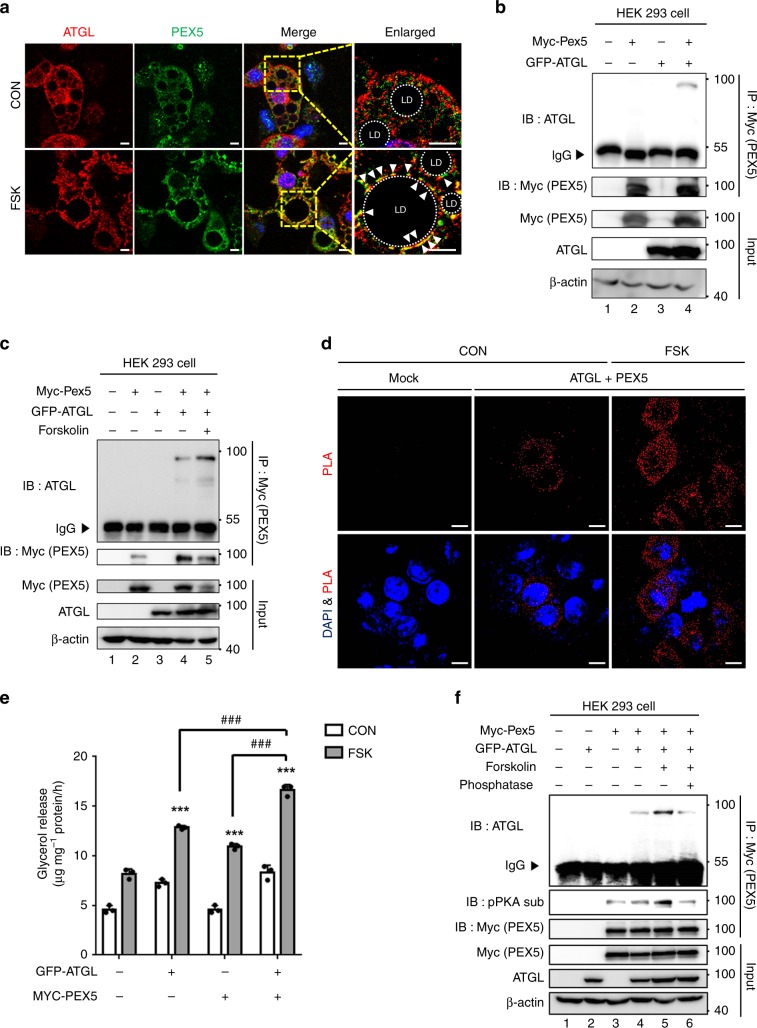
Fig. 6. PEX5 mediates stimulated lipolysis through interaction with ATGL.
a Representative images of differentiated adipocytes immunostained for endogenous ATGL (red), PEX5 (green), and DAPI (blue). Adipocytes were treated with or without FSK (10 μM) for 1 h. CON control; FSK forskolin. Scale bars, 10 μm. b, c Co-immunoprecipitation with an anti-MYC antibody and western blotting were conducted with the indicated antibodies. HEK293 cells were transfected with MYC-PEX5 and GFP-ATGL expression vectors. FSK forskolin. d In HEK293 cells, in situ proximity ligation (PLA) assay transfected with MYC-PEX5 and GFP-ATGL expression vectors without lipid challenge. FSK forskolin. Scale bars, 10 μm. e Concentrations of released glycerol from cultured COS-7 cells transfected with or without PEX5-WT and ATGL-WT expression vectors. COS-7 cells were pretreated with oleic acid (500 μM) for 48 h and FSK (25 μM) for 3 h. n = 3 for each group. CON control; FSK forskolin. Data represent the mean ± SD; ***P < 0.001 vs. Mock-CON and ###P < 0.001 group by two-way ANOVA followed by Turkey’s post-hoc test. f HEK293 cells were transfected with MYC-PEX5 and GFP-ATGL expression vectors and treated with or without FSK and phosphatase (CIAP). Co-immunoprecipitation with an anti-MYC antibody and western blotting were conducted with the indicated antibodies. IP immunoprecipitation; IB immunoblotting; IgG immunoglobulin G.

