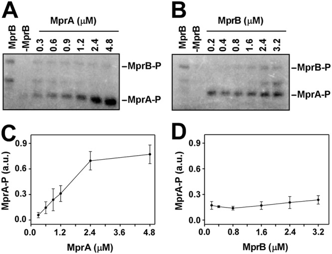
FIG 3.

In vitro phosphorylation assay using radiolabeled [γ-32P]ATP. (A) Lanes 1 and 2, autophosphorylation of 0.8 μM purified MprB (truncated) only and 2 μM MprA only, respectively; lanes 3 to 8, transfer of phosphate from 0.8 μM MprB∼P to various concentrations of MprA (0.3, 0.6, 0.9, 1.2, 2.4, and 4.8 μM). (B) Lanes 1 and 2, autophosphorylation of MprB (truncated) only and MprA only, respectively; lanes 3 to 8 transfer of a phosphate group from various concentrations of MprB (0.2, 0.4, 0.8, 1.6, 2.4, and 3.2 μM) to 0.75 μM MprA. (C and D) Average pools of MprA∼P from three replicates were plotted against concentrations of MprA and MprB, respectively. Standard errors are shown.
