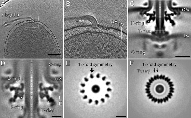
FIG 1.
In situ Vibrio flagellar motor revealed by cryo-electron tomography together with the Volta phase plate. In situ Vibrio alginolyticus flagellar motor structure obtained by cryo-ET. (A) Representative tomographic slice of a three-dimensional (3D) reconstruction of strain KK148 showing cellular features in good contrast. (B) Zoomed-in view shows the flagellar motor embedded in the outer membrane (OM) and inner membrane (IM). (C) Central slice from a subtomogram average of the intact flagellar motor shows the H ring, T ring, and C ring. (D) A central slice of the focused refined structure shows the T ring surrounding the L ring and P ring, which form the bushing part of the flagellar motor. (E) Cross-section shows a novel ring with13-fold symmetry. (F) Another cross-section shows the T ring with 26-fold symmetry. Bars, 200 nm (A), 20 nm (C), and 10 nm (D and E).

