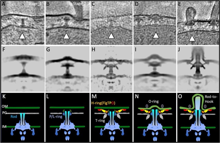
FIG 5.
Flagellar ring formations captured by time coursed cryo-electron tomography. P/L rings assemble first, a Vibrio-specific T ring composed of MotX and MotY is next, and then FlgT, FlgP, and FlgO assemble to form the H ring. O-ring formation is now ready for the rod-hook transition. (A to E) Representative slice view of 3D tomograms showing different stages (from Stage_1 to Stage_5) during the flagellar assembly. (F to J) Central section of the subtomogram-averaged structure from different stages. (K to O) Cartoon models of structures shown in panels F to J. Bar, 20 nm.

