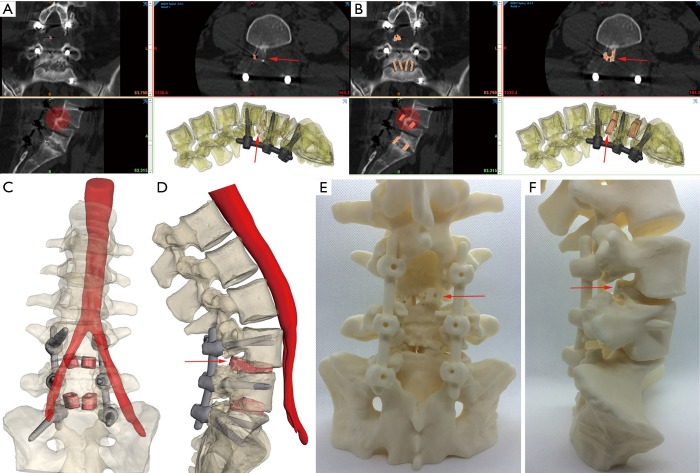
Figure 2.
Complex revision surgery planning for lumbar spine L4–L5 and L5–S1 levels using virtual and 3DP biomodelling. The patient previously had procedures using posterior approaches with the aim of stabilising and fusing L4–S1. Laminectomies had been performed with posterior pedicle rods and screws placed L4–S1 (E). Two PEEK PLIFs were positioned in each of the L4–L5 and L5–S1 interbody spaces. Fusion had not occurred at either level with the graft windows of the PEEK devices largely devoid of bone and subsidence of the L5–S1 devices visible. Loosening of the pedicle screws had resulted in an L4–L5 spondylolisthesis. In addition, the right hand side L4-L5 PLIF device had backed out from the interbody space into the spinal canal. Arrows and translucent circles show the position of the backed out PLIF. (A) PEEK implants are invisible in the CT imaging apart from embedded marker beads (shown in red); (B) shows virtual PLIF cage placement in the CT; (C) shows lumbar and sacral bone (translucent), aorta bifurcation to left and right iliac arteries, posterior rods and screws (black) virtual placement of PLIFs (light red) from an anterior viewpoint; (D) shows lateral viewpoint of the same as (C). The right hand L5 pedicle screw protrudes from the anterior surface of the L5 vertebral body (D and F) and has a trajectory coincident with the right iliac artery. Virtual reconstruction allows the distance between the L5 right hand screw tip and the iliac artery to be measured (12 mm). (E) Physical 3DP model included the reconstructed PLIF cages. The posterior viewpoint shows the result of the laminectomies as well as the right hand side L4–L5 PLIF backout. (F) 3DP model sagittal viewpoint in which the RHS backout PLIF is clearly visible, as is the protrusion of the RHS L5 pedicle screw. Revision surgery used an anterior approach to place ALIF devices. Biomodelling was used to aid with the surgical approach (vascular team) including iliac artery retractor placement with respect to the L5 screw protrusion as well as the removal of the PLIFs and insertion of the ALIF devices (neurosurgical team). The modelling showed that the spondylolisthesis at L4–L5 and large degree of lumbar lordosis kept the RHS iliac artery away from the L5 pedicle screw tip. PLIF, posterior lumbar interbody fusion; ALIF, anterior lumbar interbody fusion.

