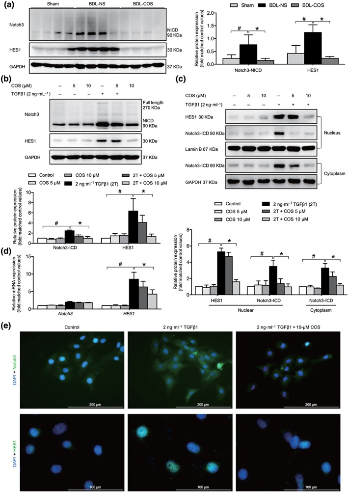
Figure 4.

Costunolide (COS) suppressed the Notch3/HES1 pathway in vitro and in vivo. COS administration down‐regulated Notch3 especially NICD3 and HES1 protein expressions in BDL livers (a) and LX‐2 cells (b). (c) The nuclear and cytoplasmic extractions of LX‐2 cells were separated after COS treatment and analysed by western blot. (d) mRNA expressions of NOTCH3 and HES1 in LX‐2 cells. The mRNA/protein expression levels were normalized against GAPDH/GAPDH. (e) LX‐2 cells in dishes were fixed in 4% paraformaldehyde, stained with DAPI (blue) and Notch3 and HES1 (green), and imaged by confocal microscopy, Scale bar: 200 μm (up panel) and 100 μm (down panel). The values are expressed as the mean ± SD of five independent assays, # P < .05, significantly different from the control group, * P < .05, significantly different from the TGFβ1 treatment group in LX‐2 cells; ANOVA followed by Tukey's test
