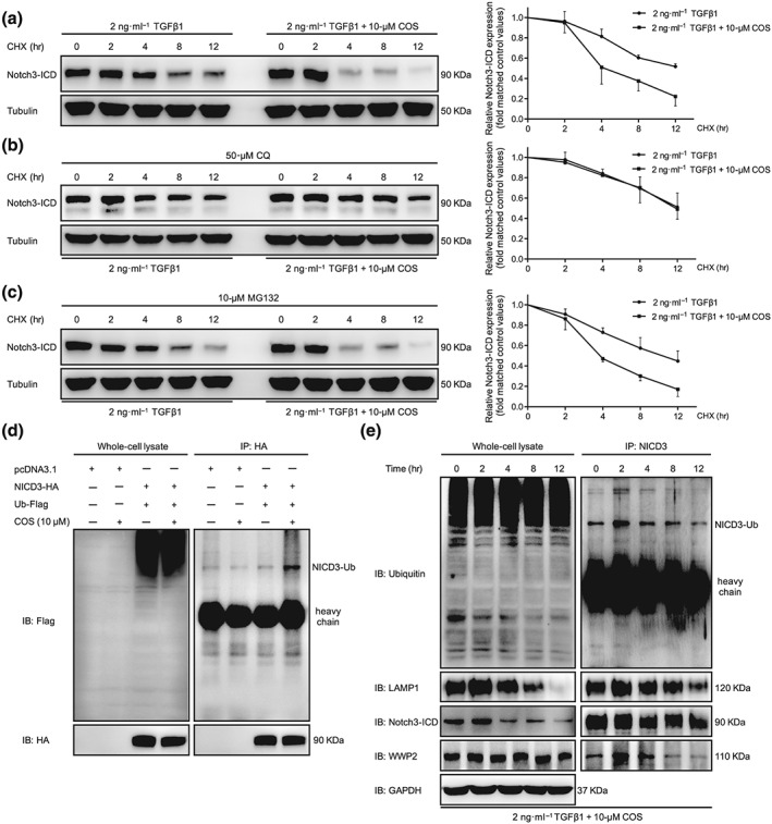
Figure 5.

Costunolide (COS) enhanced the lysosomal degradation of NICD3 in LX‐2 cells. (a) After starvation, LX‐2 cells were treated with CHX (20 mmol·L−1) and 2 ng·ml−1 TGFβ1 with or without different concentrations of COS for the indicated times. The results showed that COS inhibited the degradation of NICD3 in LX‐2 cells. (b) CQ inhibition of the lysosome blocked the degradation of NICD3 caused by COS treatment. LX‐2 cells were pretreated with CQ (100 mM) for 12 hr and then received the indicated treatments for the different times. (c) MD132 inhibition of the proteasome did not affect the NICD3 degradation. Cells were pretreated with MG132 (10 mM) for 2 hr and then received the indicated treatments for the different times. Tubulin served as a loading control. (d) NICD3‐HA was co‐transfected with Ub‐Flag into HEK293T cells. Whole cell extracts were immunoprecipitated with anti‐HA and blotted with an anti‐Flag antibody. (e) NICD3 monoubiquitination detection and the interaction among WWP2, NICD3 and LAMP1 was investigated using immunoprecipitation assays of LX‐2 cells for the indicated times. The values are expressed as the mean ± SD of five independent assays
