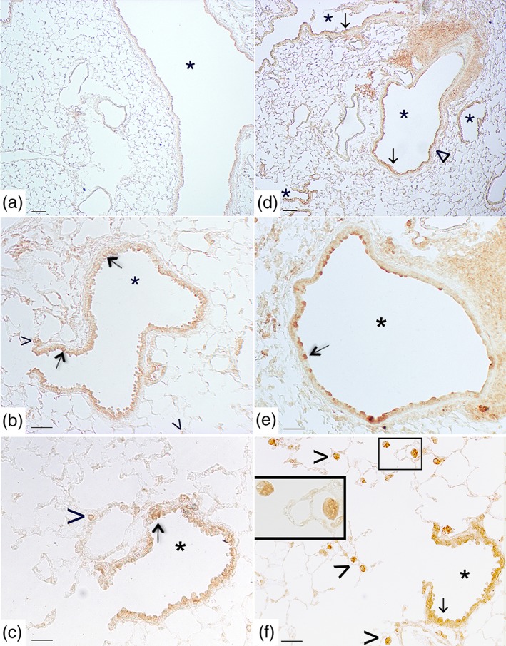
Figure 6.

Cigarette smoke induces pulmonary S1P3 up‐regulation. Panels (a), (b), and (c) representative immunohistochemical reaction for S1P3 in lung tissue from air‐exposed mice at 11 months. In panel (a), a mild positivity for S1P3 in epithelial cells of main bronchi as well as of bronchioles from air‐exposed mice is evident. In panel (b), a faint S1P3 staining is detected in epithelial cells (arrow) of small airways and in a limited number of macrophages (arrowhead). (c) A faint reaction on epithelial cells of bronchiole (arrow) and on macrophages (arrowhead) is present in air control mice. (d–f) Micrographs of lung tissue from smoking mice at 11 months. (d) The staining for S1P3 is evident and diffuse on airways epithelium (arrow) and on the smooth muscle cell layer (wedge). (e) S1P3 staining is evident on bronchial epithelial cells (arrow). (f) Epithelial cells from small airways (arrow) and macrophages (arrowhead) show a strong positivity for S1P3 in smoking mice. High magnification of two macrophages is shown in the inset. Scale bars = 100 μm (panels a and d), 60 μm (panels b and e), and 30 μm (panels c and f). Six mice for each experimental group (control and CS mice) were used
