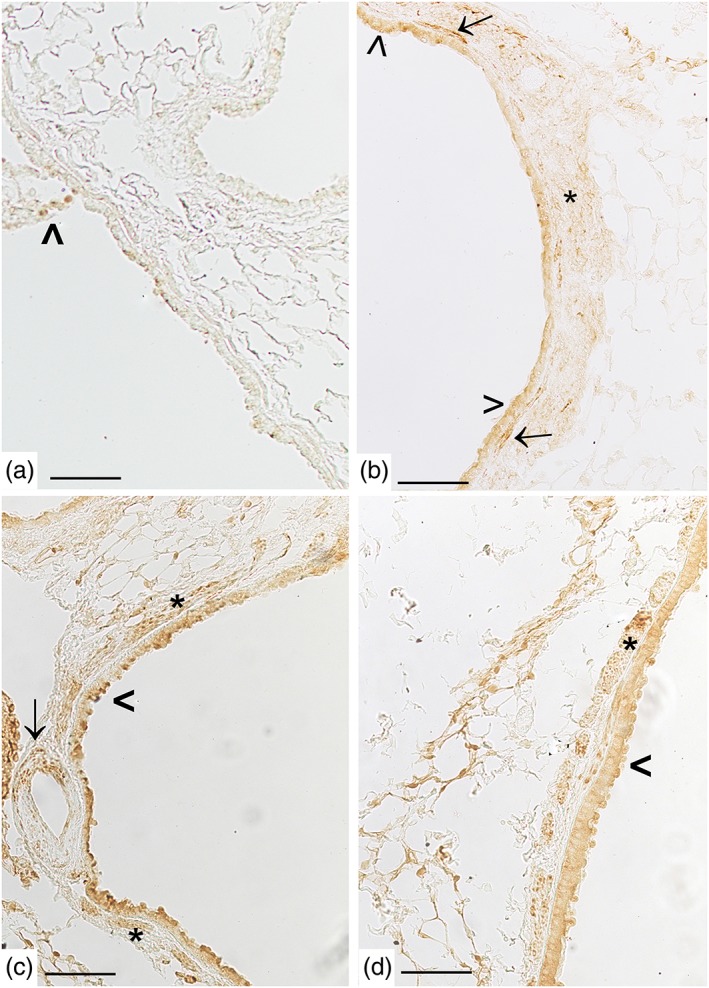
Figure 7.

Cigarette smoke induces pulmonary S1P2 up‐regulation. A mild positivity for S1P2 is present in some cells from airways epithelium from control mice (panel a, arrowhead). In panel (b), S1P2 staining is evident on epithelial cells of intraparenchymal bronchiole (arrowhead), on spindle‐shaped cells (arrow), and smooth muscle cell layer (asterisk) from mice exposed to CS for 11 months. In panel (c), S1P2 staining is evident in smooth muscle cells (asterisk), on epithelial cells of the bronchus (arrowhead), and blood vessel (arrow) from mice exposed to CS for 11 months. In panel (d), a representative lung section from a mouse exposed to CS for 9 months shows an S1P2 positivity in smooth muscle cells (asterisk). Scale bar = 100 μm. Six mice for each experimental group (control and CS mice) were used
