Figure 8.

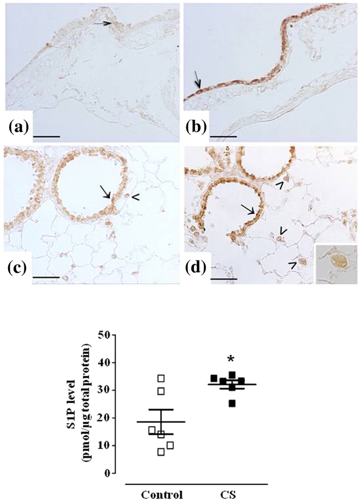
Cigarette smoke induces S1P release in the lung and a marked increase of sphingosine kinase 2. Immunostaining for Sph‐K2 in airways epithelium of main bronchi (arrow) is reported for control mice (panel a) or mice exposed to CS for 11 months (panel b). Bronchiolar epithelium (arrow) and macrophages (arrowhead) staining for Sph‐K2 is seen as a mild or strong signalling in control mice (panel c) or mice exposed to CS for 11 months (panel d), respectively. High magnification of a macrophage is shown in the inset. Scale bars = 50 μm. Six mice for each experimental group (control and CS mice) were used. S1P levels have been quantified in control and mice exposed to CS for 11 months (panel e). Data are expressed as the mean ± SEM. Seven mice for both experimental groups (control and CS mice). *P < .05 versus control
