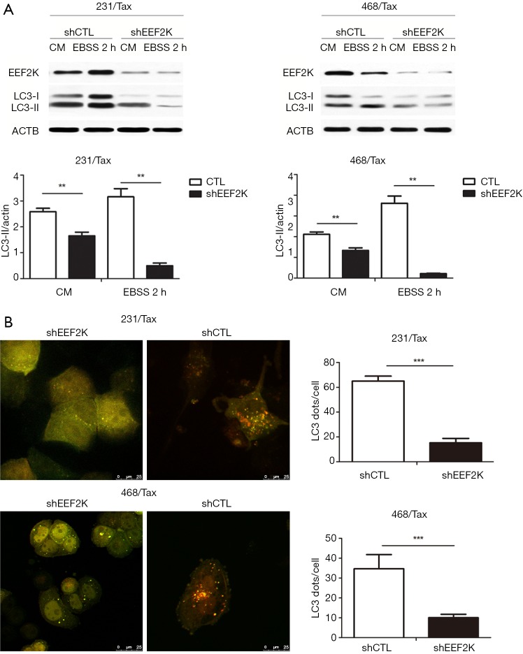
Figure 2.
Silencing eEF2K reduces autophagic flux in paclitaxel-resistant TNBC cell lines. MDA-MB-231 and MDA-MB-468 paclitaxel-resistant cells were transfected with nontargeting control shRNA (shCTL) and shRNA targeting eEF2K (sheEF2K). Silencing eEF2K markedly suppressed autophagic flux, as shown by the decreased number of LC3 dots and accumulation of LC3-II protein in eEF2K-depleted cells at baseline and after EBSS treatment. (A) Western blot analysis of LC3 I, LC3-II, and eEF2K levels in 231 resistant (231/Tax, left) and 469 resistant (468/Tax, right) cells. The results are representative of three independent experiments (Student’s t-test; **, P<0.01); (B) representative images showing the distribution of LC3 dots (autophagy marker) in eEF2K knockdown cells expressing mRFP-GFP-LC3. Cells were incubated in the presence of Baf A1 (final concentration: 100 nM). The number of GFP+/mRFP+ (yellow) dots was determined in 50 to 100 cells. The data are presented as the mean ± SD of three independent experiments (Student’s t-test; ***, P<0.001). eEF2K, eukaryotic elongation factor 2 kinase; CM, complete medium; EBSS, Earle’s balanced salt solution; TNBC, triple-negative breast cancer; mRFP, monomer red fluorescent protein; GFP, green fluorescent protein; SD, standard deviation.

