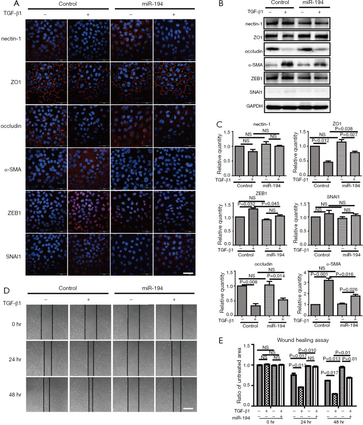
Figure 3.
miR-194 overexpression suppressed TGF-β1-induced EMT in ARPE-19 cells. (A) ARPE-19 cells transfected with empty plasmid or miR-194 were treated with 10 ng/µL TGF-β1 for 24 h. Immunofluorescence staining showing partially disrupted ZO1 staining, diminished occludin signal, and no apparent significant change to nectin-1 compared to the untreated groups; miR-194 improved or maintained ZO1 integrity and altered occludin redistribution to the membrane. α-SMA and ZEB1 in the TGF-β1 treatment group had stronger immunoreactivity compared to the untreated group; miR-194 attenuated α-SMA and ZEB1 expression patterns. (B,C) Western blotting results were consistent with that of immunofluorescence staining. (D) Wound healing assay supported the premise that miR-194 inhibited cell migration ability. NS, no significance. Scale bar =50 µm (A), 200 µm (D). The data were the mean ± SEM. Cell experiments were repeated at least 3 times. The one-way analysis of variance with Tukey’s honestly significant difference post hoc test was used to calculate the statistical significance.

