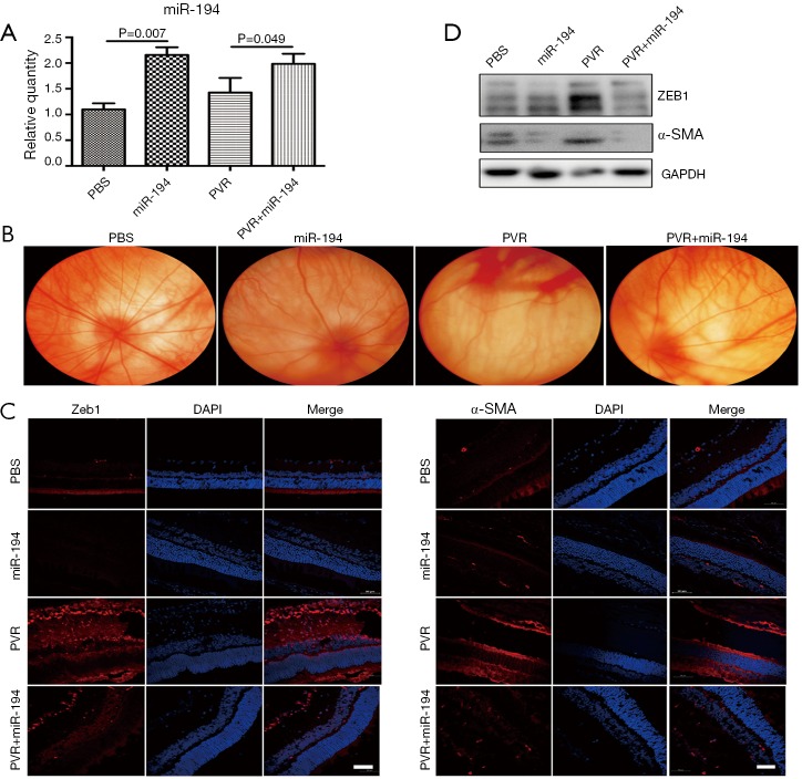
Figure 6.
Exogenous administration of agomir-194 suppressed PVR effectively in the rat model. (A) The expression of miR-194 was increased after the miR-194 intravitreal injection in the rat PVR model. (D) Western blotting showed significantly increased ZEB1 and α-SMA protein levels in PVR retina (P<0.05) compared with the PBS group, and miR-194 intervention reduced this phenomenon. (B) Fundus photography showed the typical retinal fold in the PVR group; with miR-194 intervention, the fundus appeared better, i.e., without retinal fold and with relatively straight vessels. (C) Immunofluorescence staining showed that ZEB1 and α-SMA immunoreactions were very strong in the PVR group; under miR-194 intervention, their signal was significantly attenuated, which was consistent with the Western blotting results. The data were the mean ± SEM (n=6). The one-way analysis of variance with Tukey’s honestly significant difference post hoc test was used for statistical analysis. Scale bar =50 µm.

