Fig. 4.

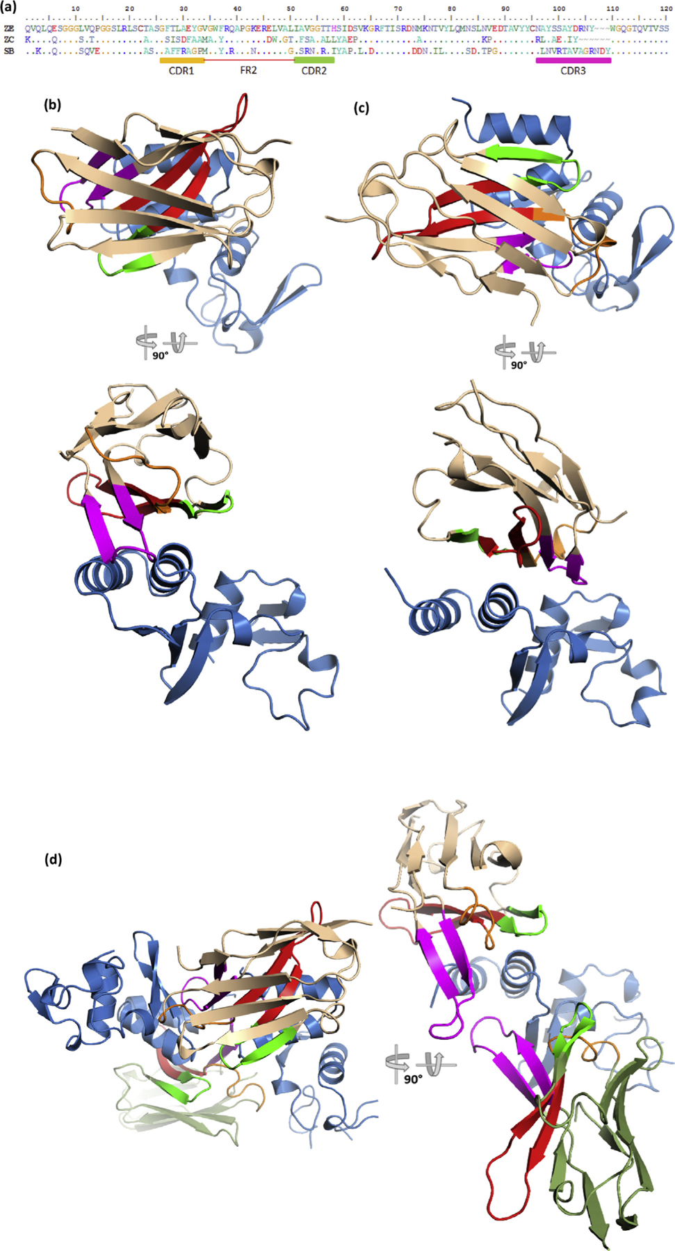
(a) Amino acid sequences of the three sdAbs under study with CDR and FR boundaries denoted and colored; CDR1, orange; CDR2, green; CDR3, magenta; FR2, red. “Top down” and “side-on” views of the antibody–antigen complexes of (b) sdAb ZE + SUDV NP634, (c) sdAb ZC + EBOZ NP634, and (d) sdAb SB + SUDV NP634. “Side-on” view of the sdAb SB complex has one of the NP634 components removed for clarity to show the long CDR3 loops of both sdAbs more clearly. Colors denoted are as follows; sdAb, wheat; NP634, sky blue; for the sdAb SB complex, the alternative sdAb is colored moss green; CDR and FR colors matches the primary structure. EBOV, Zaire ebolavirus; SUDV, Sudan ebolavirus; sdAb, single-domain antibody; FR2, framework 2.
