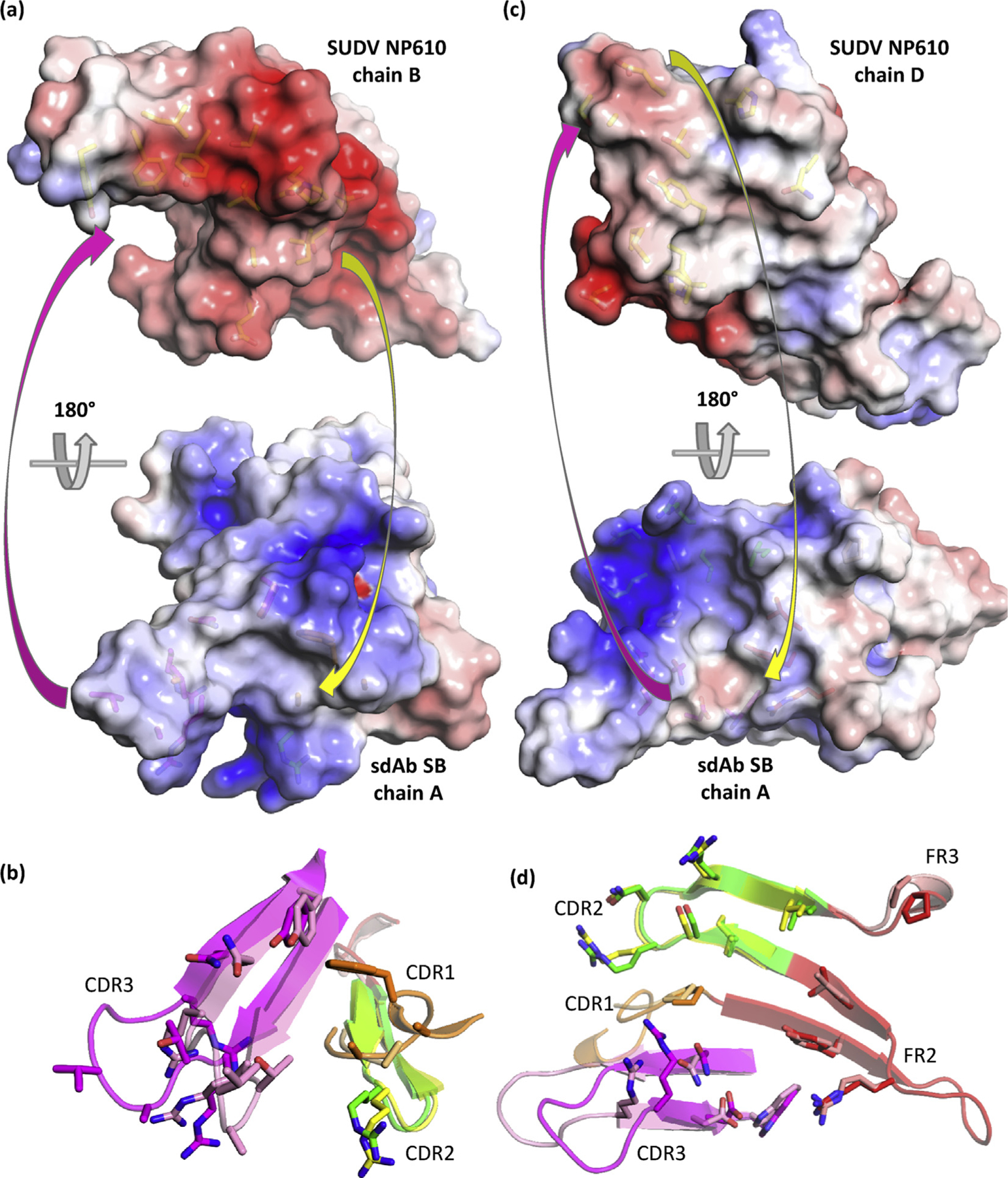
Fig. 9.

Engagement of SUDV NP610 by sdAb SB. (a) Top: antibody’s eye view of the epitope on chain B of NP610. Bottom: NP’s eye view of the sdAb paratope. Arrows indicate some of the major complementarities that are visible. (b) Overlay of the sdAb SB CDRs and FR2, aligned with the NP634’s eye view as cartoons with bound/unbound color scheme as follows: CDR1, orange/beige; CDR2, green/yellow; CDR3, magenta/pink; and FR2, red/salmon. (c) Top: antibody’s eye view of the epitope on chain D of NP610. Bottom: NP’s eye view of the sdAb paratope. Arrows indicate some of the major complementarities that are visible. (d) Overlay of the sdAb SB CDRs, FR2 and FR3, aligned with the NP634’s eye view as cartoons with bound/unbound color scheme as mentioned previously. SUDV, Sudan ebolavirus; NP, nucleoprotein; sdAb, single-domain antibody; FR2, framework 2.
