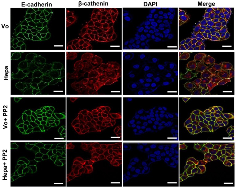
Figure 3.
Immunofluorescent staining. Control (Vo) and heparanase overexpressing T47D cells (Hepa) were left untreated or were treated with PP2 (5 μM) for 3 h. Cells were then fixed with 4% PFA, permeabilized, and subjected to immunofluorescent staining applying anti-E-cadherin (green) and anti-β-catenin (red) antibodies. Merged images are shown in the right panels together with nuclear counterstaining (blue). Shown are representative images (confocal microscopy) at ×100 magnification. Note that far more E-cadherin and β-catenin are recruited to cell-cell contacts following Src inhibition with PP2. Scale bars represent 15 microns.

