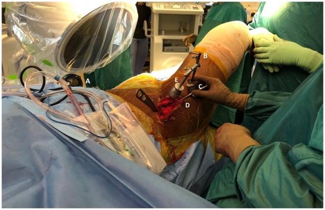
Fig. 2.

The Intellijoint HIP® 3D mini-optical navigation tool in clinical use during PAO surgery. The camera (A), enclosed in its sterile drape, is attached to the contralateral iliac crest via two screws (out of view). The tracker (B) is magnetically attached via a V-block adaptor (C) to a surgical probe (D) to allow for real-time measurement of acetabular orientation. Adjustments to acetabular fragment orientation can be made with the Schanz pin connected to a T-handle (E), with the probe and tracker used to independently confirm positioning.
