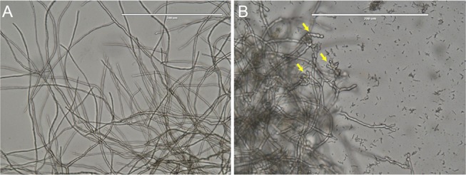
Figure 2.

Microscopic analysis. (A) Control M. phaseolina having extended straight mycelia with normal branching and septation. (B) Microscopic examination of the mycelia of M. phaseolina challenged with B. contaminans NZ at the intersection with the zone of inhibition. The arrows showing increased frequency of septa, branching, and swollen mycelia.
