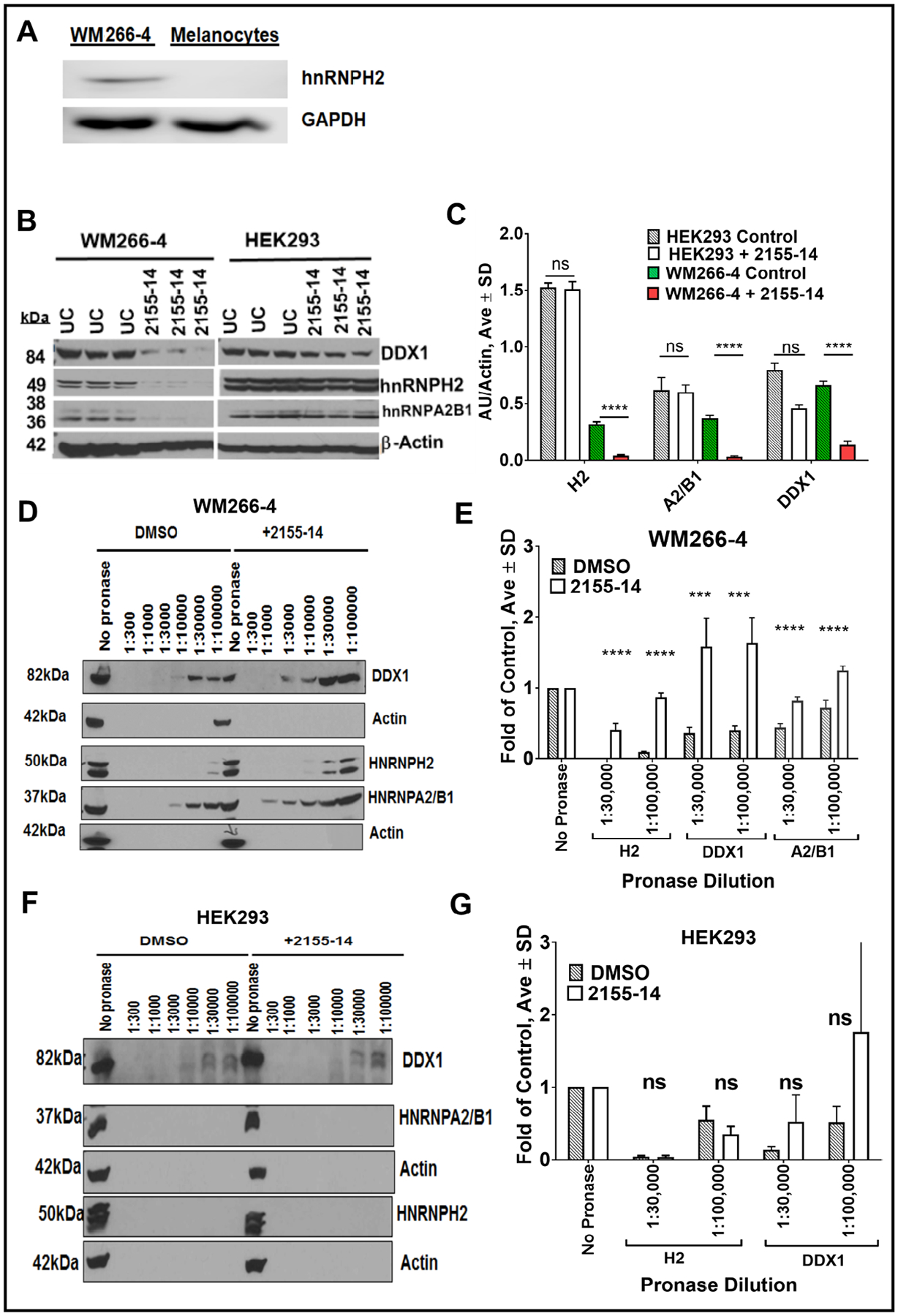
Fig. 11.

Confirmation of DDX1, hnRNP H2, and hnRNP A2/B1 as targets of 2155–14. (A) Western blot of hnRNP H2 in WM266–4 melanoma cells and melanocytes. (B) Western blot of WM266–4 melanoma and HEK293 cell lysates. Live cells were incubated in presence and absence of 2155–14. (C) Quantification of western blots for (B). (D) Western blot of DDX1, hnRNP H2, and hnRNP A2/B1 in WM266–4 cell lysates after digestion with pronase in the presence and absence of 2155–14. (E) Quantification of western blots for (D). (F) Western blot of DDX1, hnRNP H2, and hnRNP A2/B1 in HEK293 cell lysates after digestion with pronase in the presence and absence of 2155–14. (E) Quantification of western blots for (F). One-way analysis of variance (ANOVA) was used followed by Dunnett post hoc test. The data shown were the mean ± SD, n=3. ***** - p<0.0001, *** - p<0.001, ** - p<0.01, * - p<0.05, rest = no significance.
