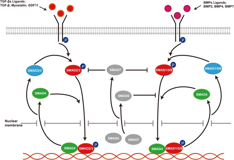
Fig. 1.
TGF-β/SMAD signaling. TGF-βs ligands such as TGF-β, Myostatin, and GDF11 in TGF-β/SMAD signaling bind to cell membrane receptors to phosphorylate the intracellular downstream SMAD2/3 (R-SMADs), and BMPs ligands such as BMP2, BMP4, and BMP7 phosphorylate the SMAD1/5/8 (R-SMADs). Activated R-SMADs form a complex with SMAD4 and then translocates to the nucleus to regulate the expression of specific genes. After the genes respond to the TGF-β/SMAD signaling, the R-SMADs–SMAD4 complex in the nucleus is depolymerized and they re-enter the cytoplasm. I-SMADs comprise SMAD6 and SMAD7, which negatively regulate TGF-β/SMAD signaling. In the resting state, I-SMADs mainly tend to be in the nucleus. Transcriptionally activated by TGF-β/SMAD signaling, SMAD7 shuttling from the nucleus to the cytoplasm prevents R-SMAD phosphorylation. SMAD6 competes with SMAD1 for binding to SMAD4

