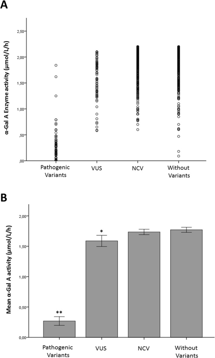
Fig. 1.
Enzymatic profile of GLA genotypes. (a) Scatter plot of the α-Gal A activity distribution in males with FD suspicion in different groups. The figure shows that most males with VUS, NCV and without variants present α-Gal A levels above 1 μmol/L/h, while patients with pathogenic variants presented α-Gal A levels lower than 1 μmol/L/h. Some outliers were found in each group. Three patients with pathogenic variants presented enzyme activity above 1 μmol/L/h, while twenty-four patients with only non-coding variants, twenty without variants and seven with VUS, being four with A143T, two with D313Y and one with R356Q, presented enzyme activity below 1 μmol/L/h. (b) Correlation analysis between α-Gal A level in DBS and GLA genotypes. The graphic shows the mean enzymatic activity detected in males in all the GLA variant groups. The data are expressed as mean ± S.E.M. **P < 0.001 known pathogenic mutation (0.27 μmol/L/h ± 0.03, N = 83) versus VUS (1.58 μmol/L/h ± 0.04, N = 76), non-coding variants (1.73 μmol/L/h ± 0.02, N = 289) and the group without variants in GLA (1.77 μmol/L/h ± 0.02, N = 335); *P = 0.013 VUS versus NCV and *P = 0.01 VUS versus patients without variants

