Fig. 3.
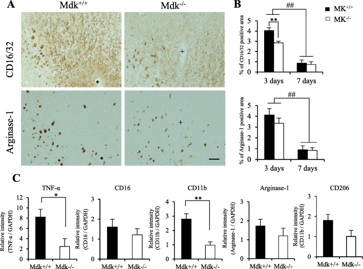
Effect of MK-deficiency on M1 and M2 microglia/macrophages phenotype marker after TBI. The M1 and M2 phenotype markers (CD16/32 and arginase-1, respectively) were expressed in the perilesional site of Mdk+/+ and Mdk−/− mice at 3 days (a). The immunohistochemical staining was performed through serial sections of mice (corresponding to * and + in a). The ratios of the CD16/32-immunoreactive area were significantly reduced in Mdk−/− mice compared to Mdk+/+ mice at 3 days. The CD16/32- and arginase-1-immunoreactive areas were significantly decreased at 7 days (b). RT-qPCR analysis revealed the mRNA levels of the M1 phenotype markers (TNF-α, CD11b) to be significantly downregulated in Mdk−/−than in Mdk+/+ mice (c). Data are presented as mean ± SE (n = 5 mice/group in immunohistochemistry, n = 3–4 mice/group in RT-qPCR). *p < 0.05, **p < 0.01 (comparison with MK+/+ and Mdk−/−). ##p < 0.01 (comparison with 3 days and 7 days). Scale bar = 50 μm (all panels)

