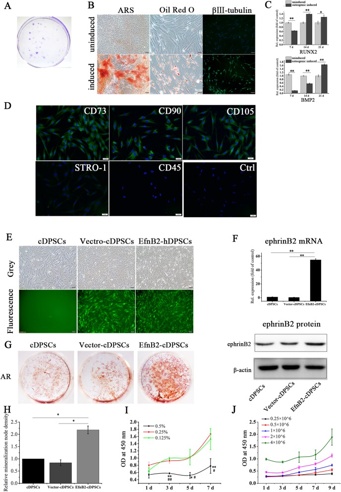
Fig. 5.
Culture, characterization, and transfection of cDPSCs and proliferation of cDPSCs in PuraMatrix. a Cell colonies stained with crystal violet. b, c Verification of osteogenic, adipogenic, and neurogenic differentiation capabilities of cDPSCs. Scale bar of left and right images, 100 μm; scale bar of middle images, 50 μm. *p < 0.05 and **p < 0.01. d Stem cell markers of cDPSCs. Scale bar = 1 mm. e, f Verification of green fluorescence expression and ephrinB2 upregulation in transfected cDPSCs. Scale bar = 100 μm. **p < 0.01. g Alizarin Red S staining of cDPSCs, Vector-cDPSCs (ephrinB2 overexpression control), and EfnB2-cDPSCs (ephrinB2 overexpression) on day 24 of osteogenesis. h Alizarin Red S staining intensity was quantified with ImageJ. *p < 0.05. i Proliferation of cDPSCs (1 × 10⁶ cells/ml) in 0.5%, 0.25%, and 0.125% PuraMatrix. *p < 0.05 and **p < 0.01 vs. 0.25% PuraMatrix; # p < 0.05 and ## p < 0.01 vs. 0.125% PuraMatrix. j Proliferation of cDPSCs at different cell densities (0.25, 0.5, 1, 2, or 4 × 10⁶ cells/ml) in 0.25% PuraMatrix. Data are shown as mean ± SD. Assays were repeated three times

