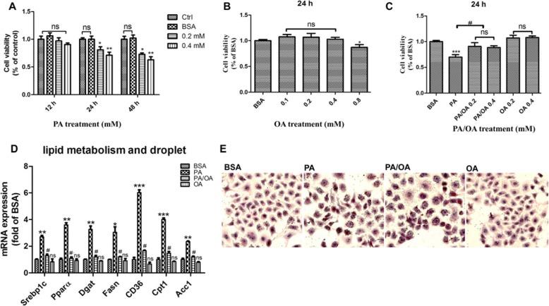
Fig. 1.
Oleic acid protected HepG2 cells from palmitic acid induced Lipotoxicity. Viability of HepG2 cells was assessed using the CCK8 assay. a. and b. Alternatively, cells were treated with PA or OA alone for 12 h,24 h or 48 h. c. Cells were concomitantly incubated with PA and OA for 24 h. d. HepG2 were treated with 0.4 mM PA, 0.2 mM OA or combination of 0.4 mM PA plus 0.2 mM OA (PA/OA). The mRNA expression of key genes governing lipid metabolism were detected after 24 h treatment, and β-ACTIN was used as an internal control; e. Cells were stained with Oil Red O and lipid accumulation was visualized under a microscope at 200 × magnification after 24 h treatment. The data are presented as means ± SD for 3–5 biological replicates; *P < 0.05, **P < 0.01, *** P < 0.001vs. BSA;#P < 0.05,vs. PA; ns no significant differences between two connected groups

