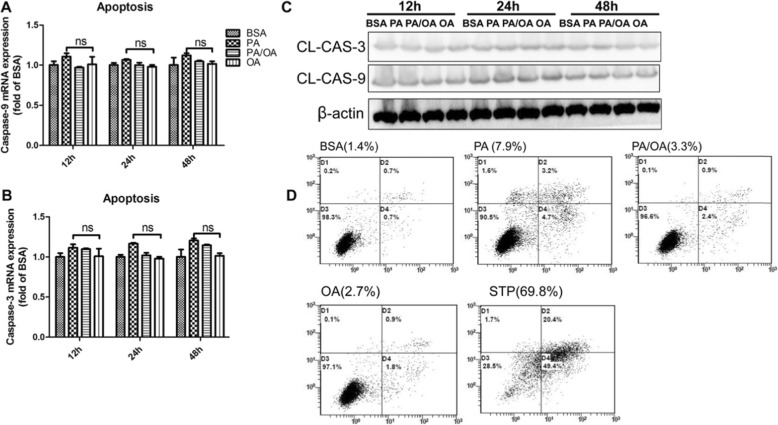
Fig. 2.
Apoptosis is not the main form of cell death caused by palmitic acid induced Lipotoxicity. HepG2 were treated with 0.4 mM PA, 0.2 mM OA or combination 0.4 mM PA plus 0.2 mM OA (PA/OA). a and b. The mRNA expression of key genes governing apoptosis were detected after 24 h treatment, and β-ACTIN was used as an internal control. c. Representative western blots of Cleaved-caspase3/9 after 24 h treatment, and β-ACTIN was used as a protein-loading control. d. Apoptosis assay using FCM with AV/PI staining after 24 h treatment. The numbers at the lower or upper right indicate the percentage of sum of early and late apoptotic cells. The data are presented as means ± SD for 3–5 biological replicates; *P < 0.05, **P < 0.01, ***, P < 0.001vs. BSA; ns no significant differences between two connected groups

