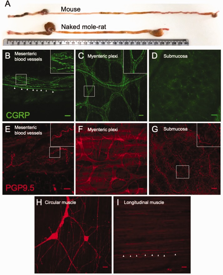
Figure 1.
Extrinsic sensory innervation of NMR colorectum. (a) Comparison of mouse and NMR gastrointestinal tracts from anus (left) to oesophagus (right), with a 30-cm ruler providing scale. Whole-mount immunostaining for CGRP in the mesentery (b; inset, nerve fibres encircling a mesenteric blood vessel. Arrows, example nerve fibre on the blood vessel margin), myenteric plexi (c; inset, extrinsic nerve fibres infiltrating myenteric ganglia) and submucosa (d) of NMR. Equivalent nerve fibre staining was also observed with PGP9.5 in the mesentery (e; inset, nerve fibre surrounding mesenteric blood vessel), myenteric plexi (f), submucosa (g; inset, nerve fibre surrounding the base of a mucosal villi), circular muscle (h) and longitudinal muscle (i; arrows, nerve fibre innervating longitudinal muscle). Scale bar in each panel: 50 µm. CGRP: calcitonin gene-related peptide; PGP9.5: protein gene product 9.5.

