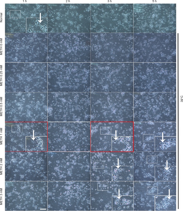
Figure 1.
Morphological effect of METH + 39°C on cortical neurons.
We applied 0, 0.25, 0.5, 1, 2, 3 mM of METH + 39°C for 1, 2, 3 and 5 hours in the experiment. The neuronal cells are photographed under the inverted microscope. The drug concentration and duration times in the red square boxes are our selected parameters for later experiments. The white arrows show morphological changes of cell necrosis. The larger picture in the lower right corner of panels is the enlargement of the smaller picture in the corresponding picture. Scale bar: 50 μm in all the panels. The baseline in each of the 100 μm in all enlarged pictures in the lower right corners of panels equals 100 μm. METH: Methamphetamine.

