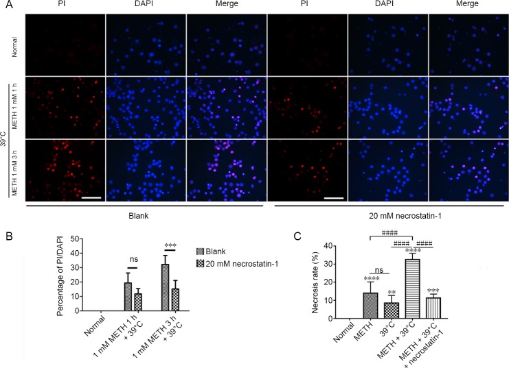
Figure 2.
Necrosis of rat cortical neurons induced by METH + 39°C.
(A) PI staining for cortical neurons by METH + 39°C, left. Pretreatment with 20 mM necrostatin-1, right; scale bars: 100 μm. (B) Statistics of PI uptake rate (mean ± SD, n = 5; two-way analysis of variance followed by the Bonferroni post hoc test), ***P < 0.001. (C) Necrosis rate of cortical neurons detected by lactate dehydrogenase assay after METH + 39°C for 3 hours, with and without pretreatment with necrostatin-1 (mean ± SD, n = 6, one-way analysis of variance followed by Tukey’s multiple comparison test). **P < 0.01, ***P < 0.001, ****P < 0.0001, vs. normal group; ####P < 0.0001. METH: Methamphetamine; ns: not significant; PI: propidium iodide.

