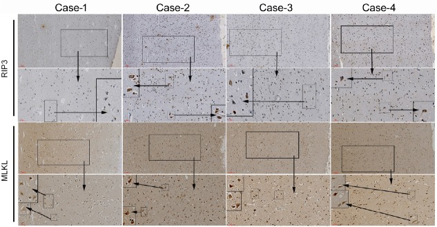
Figure 5.
Immunofluorescence staining of human cadaver frontal cortex sections.
The causes of death are listed in the Table 1. RIP3 and MLKL are stained by immunohistochemistry for each case. The small- to medium-sized frame is a local 4 × magnification of the larger one. Scale bars: 100 μm in row one and three, 50 μm in row two and four. MLKL: Mixed lineage kinase domain-like protein; RIP3: receptor-interacting protein 3.

