
Figure 5.
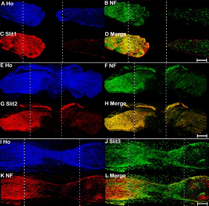
Double immunohistochemical staining of Slit1–3 and NF in mouse sciatic nerve at 7 days after transection injury.
(A–D) Double staining of Slit1 (red, Alexa Fluor 568) and NF (green, Alexa Fluor 488) in a 7-day post-transection mouse sciatic nerve showing Slit1 expression in regenerating axons of the proximal nerve stump. (E–H) Double staining of Slit2 (red, Alexa Fluor 568) and NF (green, Alexa Fluor 488) in a 7-day post-transection mouse sciatic nerve showing Slit2 expression in the proximal nerve stump. (I–L) Double staining of Slit3 (green, Alexa Fluor 488) and NF (red, Alexa Fluor 568) in a 7-day post-transection mouse sciatic nerve showing Slit3 expression in the proximal nerve stump, nerve bridge, and distal nerve stump. The proximal nerve stump is on the left, the distal nerve stump is on the right, and the nerve bridge is indicated between the two dashed lines. Scale bars: 400 μm. Ho: Hoechst 33342; NF: Neurofilament heavy chain; Robo: Roundabout.