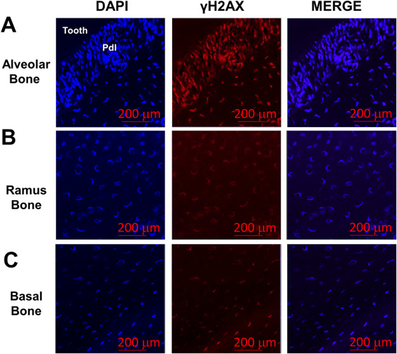
Fig. 2.

Levels of DNA damage are increased in alveolar bone osteocytes. (a) Presence of γH2AX, a marker of DNA damage, was evaluated by immunofluorescence in sections from alveolar bone from 6 month-old WT female mice in vivo. Stronger γH2AX (red signal) was observed both within the periodontal ligament (Pdl) and in neighboring alveolar osteocytes. Ramus (b) and underlying basal bone (c) displayed lower amounts of γH2AX signal. Cell nuclei were labeled with DAPI (blue signal). The red bars denote the magnification scale (200 μm).
