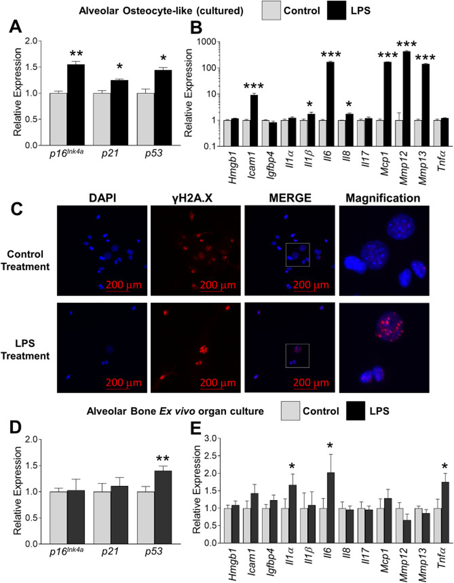
Fig. 3.

Chronic LPS exposure increases several indices of cellular senescence in alveolar bone osteocytes. (a-b) Alveolar bone osteocyte-like cells were prepared from 6 month-old WT female mice, treated with either vehicle control or P. gingivalis LPS (10 ng/ml) for six days and assayed for expression of selected senescent marker and SASP genes using QPCR. Data represent Mean ± SEM (n=10). *P ≤ 0.05, **P ≤ 0.01 and ***P ≤ 0.001 relative to vehicle control. (c) Alveolar osteocyte-like cells were treated with control or LPS as in panels a-b and stained with an antibody against γH2AX (red signal). The boxed inset in the Merge panel is shown enlarged in the Magnification panel to observe single γH2AX-stained foci. Cell nuclei were labeled with DAPI (blue signal). The red bars denote the magnification scale (200 μm). (d-e) Intact (non-digested) left and right alveolar bone blocks were treated with vehicle control or LPS, respectively, for 6 days as assayed for expression of selected senescent marker and SASP genes using QPCR. Data represent Mean ± SEM (n=6). *P ≤ 0.05, and **P ≤ 0.01 relative to vehicle control.
