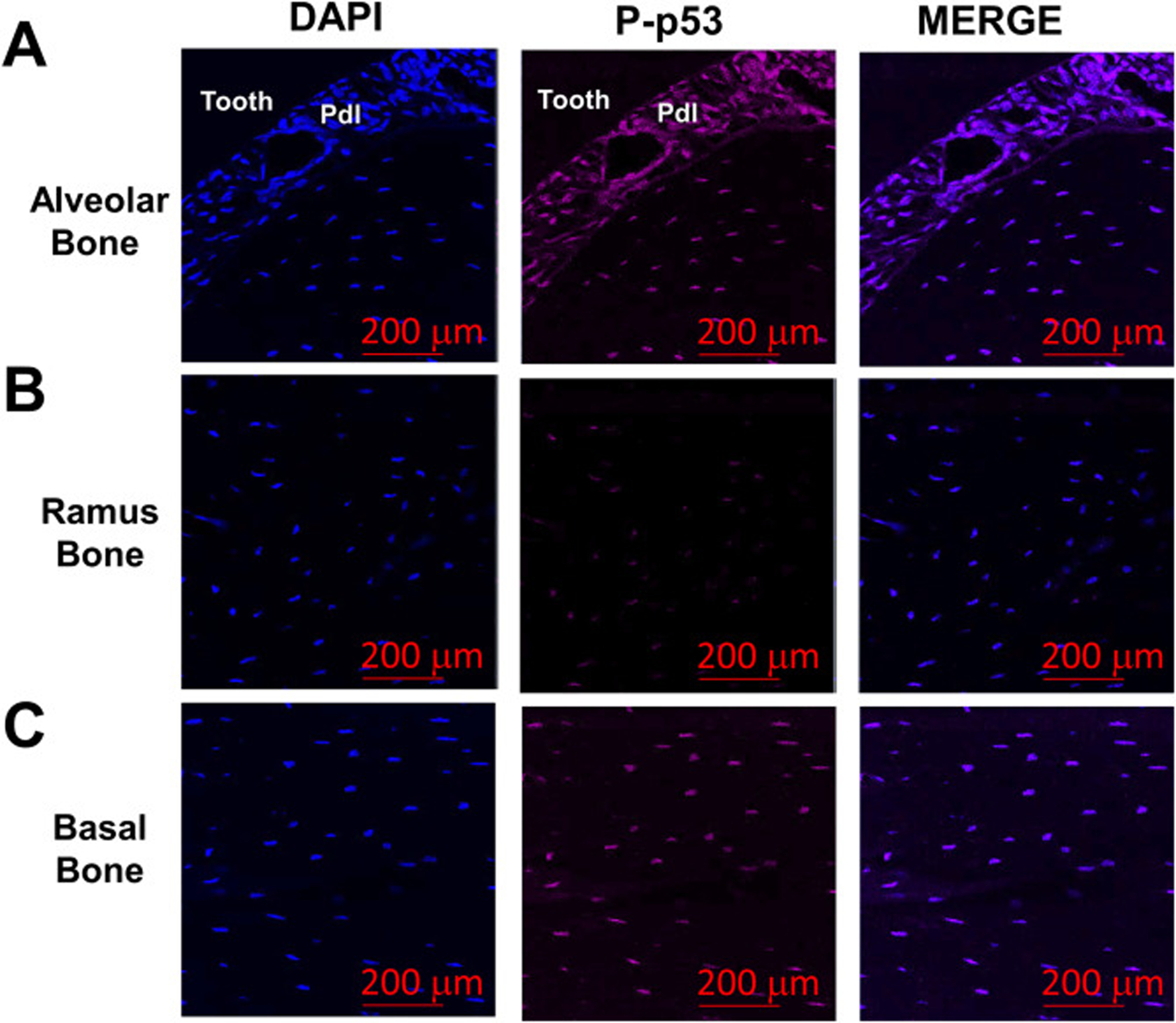
Fig. 4.

P-p53 protein levels are increased in alveolar bone osteocytes in vivo. (a-c) Bone sections from alveolar, ramus and underlying basal bone from 6 month-old WT female mice were stained for Phospho(P)-p53 levels (magenta signal). Cell nuclei were labeled with DAPI (blue signal). Cell nuclei were labeled with DAPI (blue signal). The red bars denote the magnification scale (200 μm).
