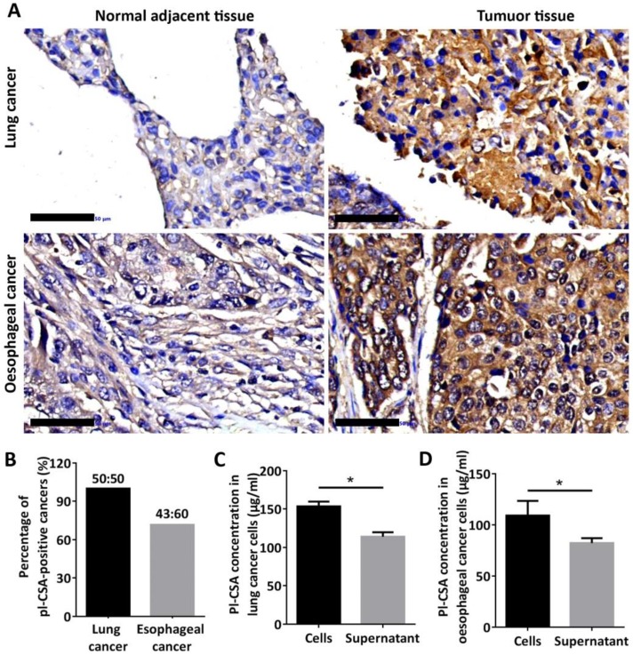
Figure 6.
Pl-CSA was synthesized in cancer tissue and cancer cell lines and released in bio-fluids. (A) Pl-CSA was detectable by immunohistochemistry in oesophageal and lung cancer tissue but not in normal adjacent tissue (scale bar: 50 μm). (B) The analysis of pl-CSA-positive cases indicated 72% and 100% positivity in oesophageal and lung cancer tissues, respectively, compared with normal adjacent tissue. (C) A small amount of pl-CSA was released into the supernatant by the lung cancer cell lines, and the pl-CSA content was significantly higher in the lysates than in the supernatants. (D) A small amount of pl-CSA was released into the supernatant by the oesophageal cancer cell lines, and the pl-CSA content in the lysates was significantly higher than that in the supernatants.

