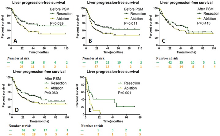
Figure 5. LPFS stratified by the size of liver metastases between the resection and ablation groups.
LPFS was significantly different before PSM for ≤ 3 cm solitary (A) and multiple lesions (B). After PSM, LPFS was not significantly different for ≤ 3 cm solitary (C) and multiple lesions (D), but was significantly longer in the resection group for >3 cm lesion (E). LPFS Liver progression-free survival, PSM Propensity score matching.

