Figure 1.

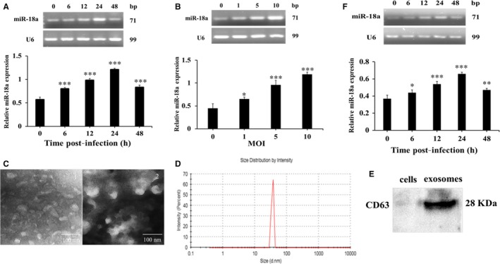
The expression of miR‐18a was induced after Mtb infection. A, The expression of miR‐18a at different time‐points in RAW264.7 macrophages infected by Mtb. B, The expression of miR‐18a at different MOI in RAW 264.7 macrophages infected by Mtb. C, Observation of exosomal characteristic by TEM. D, Estimation of the exosomal diameter by particle size analysis. E, Detection of the exosomal protein marker CD63 by Western blot. F, The expression of miR‐18a at different time‐points in exosomes derived from Mtb‐infected RAW264.7 macrophages. Data represent the means ± SD from three independent experiments. *P < .05, **P < .01, ***P < .001. Relative miR‐18a expression, miR‐18a/ internal reference U6
