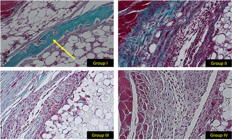
FIGURE 3.
20× histology images stained with MT. The histological slices represent the same area of each muscle pouch at the interface between the exterior of the HB scaffold and native muscle. In group I there is evidence of woven bone formation (yellow arrow). There is no evidence of bone formation in groups II–IV.

