
Figure 2.
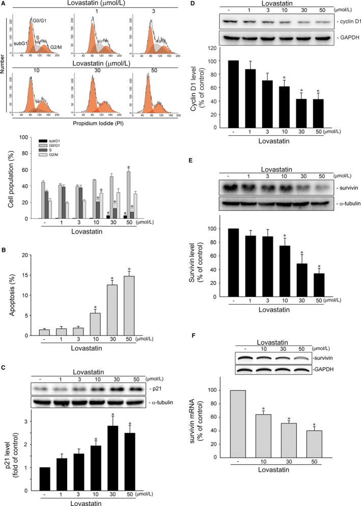
Lovastatin caused survivin reduction and apoptosis in MCF‐7 cells. A, Cells were treated with vehicle or lovastatin at indicated concentrations for 24 h. The percentage of propidium iodide‐stained cells in subG1, G0/G1, S and G2/M phases was analysed by flow cytometry as described in the ‘Materials and Methods’ section. Each column represents the mean ± SEM of four independent experiments (Statistically significant differences were determined using one‐way ANOVA, with Tukey's post hoc test. *P < .05, compared with the control group). B, Cells were treated as described in (A) for 48 h. The percentage of propidium iodide‐stained cells in apoptosis (subG1) region was analysed by flow cytometry as described in the ‘Materials and Methods’ section. Each column represents the mean ± SEM of four independent experiments (Statistically significant differences were determined using one‐way ANOVA, with Tukey's post hoc test. *P < .05, compared with the control group). MCF‐7 cells were treated with vehicle or lovastatin at indicated concentrations for 24 h. Protein levels of p21 (MW 21 kD) (C), cycin D1 (MW 36 kD) (D), survivin (MW 16 kD) (E), α‐tubulin (MW 52 kD) and GAPDH (MW 37 kD) were determined by immunoblotting. Each column represents the mean ± SEM of eight independent experiments (Statistically significant differences were determined using the Kruskal‐Wallis test. *P < .05, compared with the control group). F, Cells were treated with vehicle or lovastatin at indicated concentrations for 6 h. The survivin mRNA level was determined by an RT‐PCR as described in the ‘Materials and Methods’ section. Each column represents the mean ± SEM of three independent experiments (Statistically significant differences were determined using the Mann‐Whitney test. *P < .05, compared with the control group)