
Figure 6.
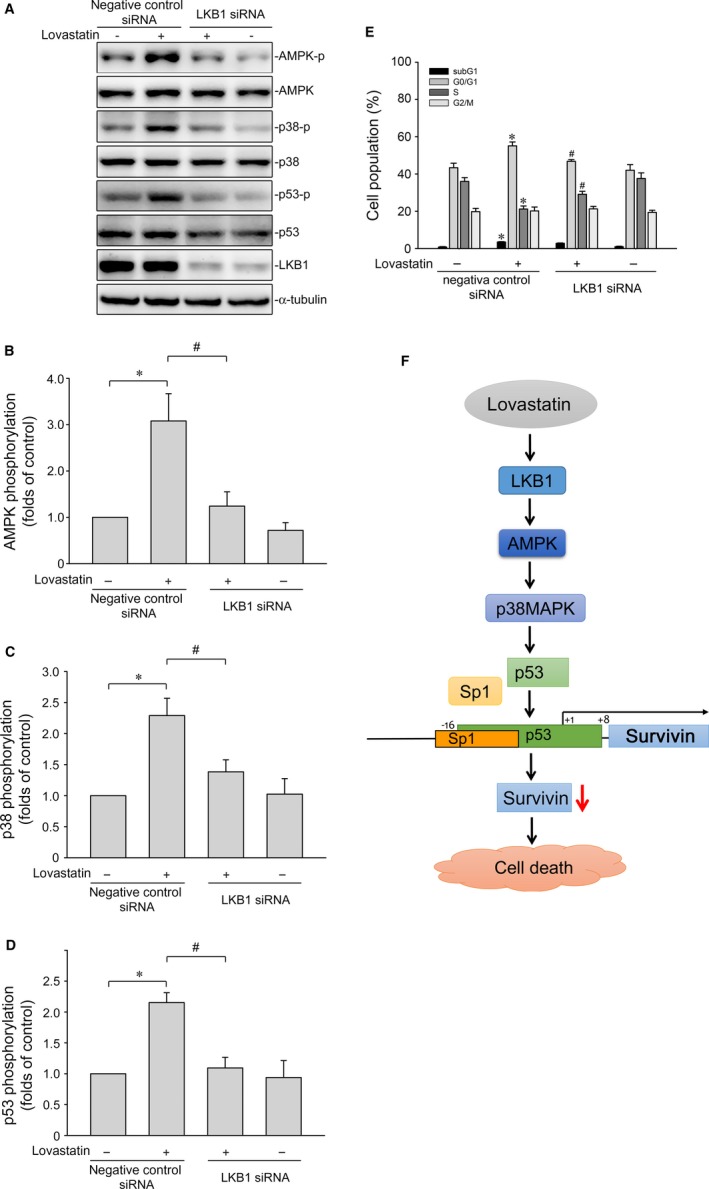
LKB1 contributes to lovastatin‐induced AMPK, p38MAPK and p53 phosphorylation in MCF‐7 cells. A, Cells were transfected with negative control siRNA or LKB1 siRNA for 48 h. After transfection, cells were treated with vehicle or lovastatin (30 μmol/L) for another 1 h. The extent of LKB1 and phosphorylation status of AMPK, p38MAPK or p53 was determined by immunoblotting. The compiled results of AMPK (B), p38MAPK (C) and p53 (D) phosphorylations are shown. Each column represents the mean ± SEM of six independent experiments (Statistically significant differences were determined using the Mann‐Whitney test. *P < .05, compared with the vehicle‐treated control group; # P < .05, compared with the group treated with lovastatin alone). (E) After transfection as described in (A), cells were treated with vehicle or lovastatin (30 μmol/L) for another 24 h. The percentage of propidium iodide‐stained cells in subG1, G0/G1, S and G2/M phases was analysed by flow cytometry. Each column represents the mean ± SEM of eight independent experiments (Statistically significant differences were determined using one‐way ANOVA, with Tukey's post hoc test. *P < .05, compared with the negative control siRNA‐transfected group; # P < .05, compared with the negative control siRNA‐transfected group in the presence of lovastatin). F, Schematic summary of the signalling pathway involved in lovastatin‐induced MCF‐7 breast cancer cell death