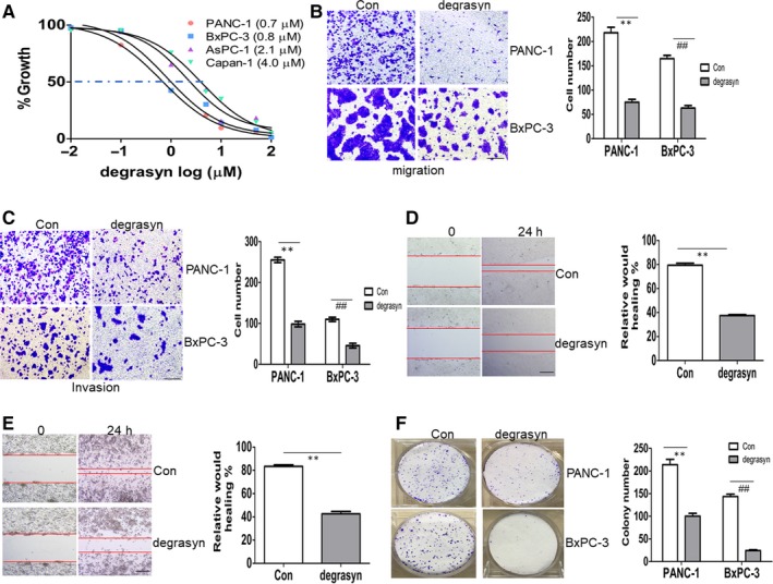
Figure 1.

The anti‐cancer ability of degrasyn in pancreatic cancer cells. (A) Four pancreatic cancer cell lines were treated with different concentrations of degrasyn for 24 hours. Cell growth was measured by CCK‐8 assay. A 50% inhibitory concentration (IC50) of degrasyn was calculated for the four pancreatic cancer cell lines. (B and C) Transwell migration (B) and invasion (C) assays were performed in PANC‐1 and BxPC‐3 cells treated with or without 1 μM degrasyn for 24 hours. ** and ## P < .01. Shown is the representative plot (Left) and summary of cell number (Right). (D and E) Wound‐healing assay was performed in PANC‐1 (D) and BxPC‐3 cells (E) treated with or without 1 μM degrasyn. ** and ## P < .01. Shown is the representative picture (Left) and summary of relative would healing (Right). (F) Colony formation was performed by methylene blue staining in PANC‐1 and BxPC‐3 cells treated with or without 1 μM degrasyn. ** and ## P < .01. Shown is the representative plot (Left) and summary of colony number (Right)
